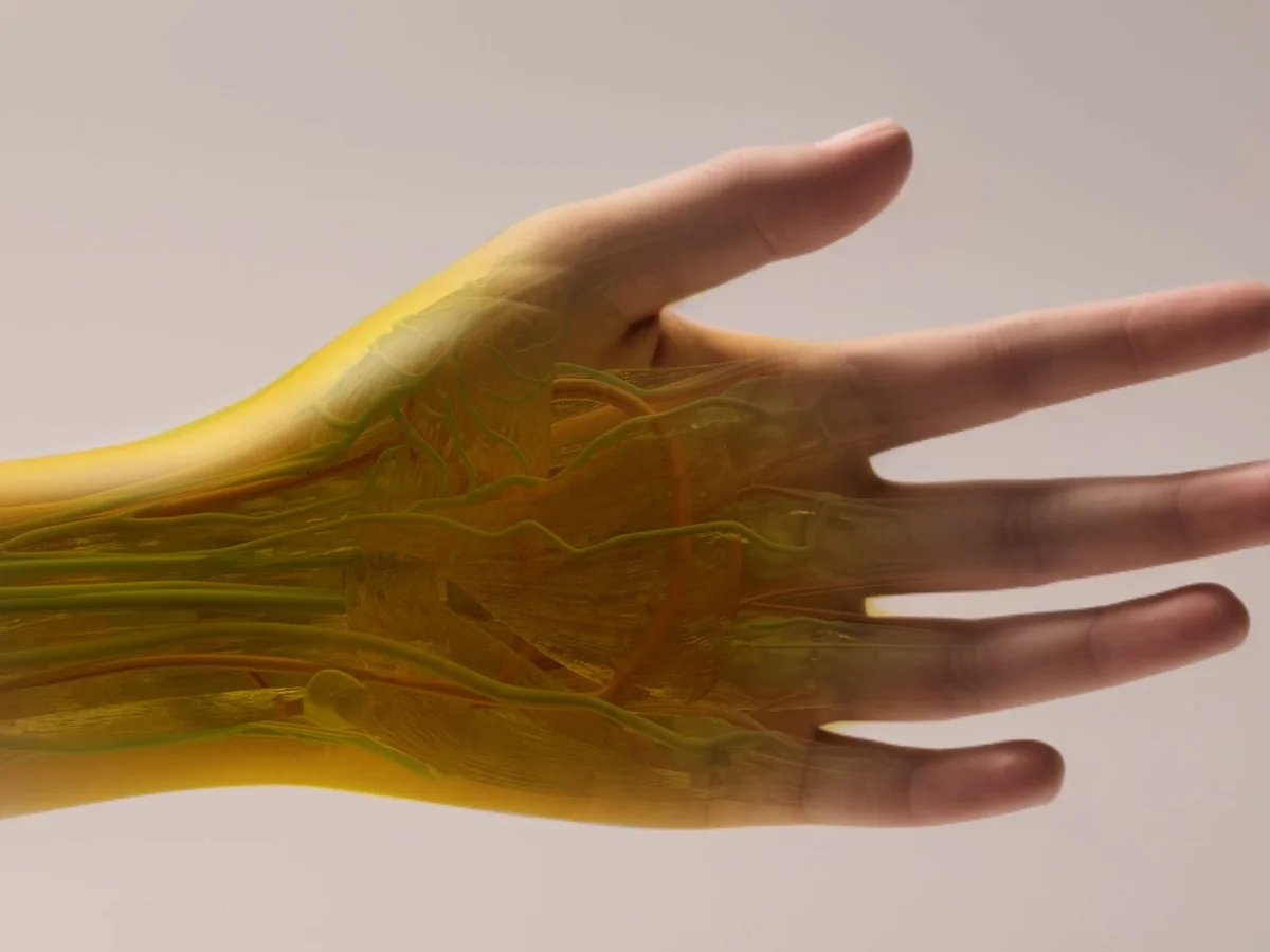
Researchers have made a significant discovery by using a common food dye to render the skin, muscles, and connective tissues of living animals temporarily transparent, enabling them to observe internal organs in real-time.
This breakthrough, made by scientists at Stanford University, reveals how the dye can be applied to animal skin to provide a clear view of underlying tissues.
For example, applying the dye to a mouse’s abdomen allowed researchers to see its liver, intestines, and bladder through the skin. Similarly, when applied to the scalp, the dye exposed the blood vessels in the brain.
Remarkably, the skin returned to its natural color once the dye was washed off, suggesting a wide range of potential applications for humans.
The researchers believe this technique could be useful in locating injuries, finding veins for blood draws, diagnosing digestive disorders, and even detecting tumors without the need for invasive procedures.
Dr. Guosong Hong, a senior researcher on the project, commented on the potential benefits: “Doctors might soon be able to diagnose deep-seated tumors by simply examining a person’s tissue without needing to remove it surgically.
It could also make blood draws less painful by helping phlebotomists locate veins under the skin more easily.”
The technique draws parallels with the story of Griffin in HG Wells’ The Invisible Man, where the character discovers that invisibility can be achieved by matching an object’s refractive index to the surrounding air.
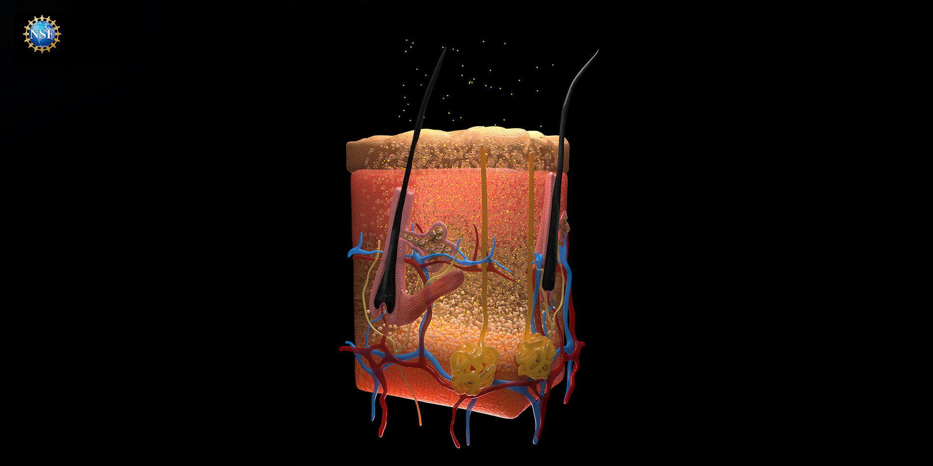
Similarly, biological tissues scatter light due to differences in refractive indices between structures such as cell membranes and nuclei.
This scattering causes tissue to appear opaque, much like how a pencil appears bent when submerged in water.
Dr. Zihao Ou and his colleagues at Stanford theorized that specific dyes might alter these refractive indices and allow certain wavelengths of light to pass more easily through biological tissues.
The dyes change the refractive index of tissues that absorb them, reducing light scattering and making the tissue more transparent.
In experiments described, the researchers demonstrated the effects of a yellow food dye called tartrazine, commonly found in US products like Doritos and SunnyD, on a fresh chicken breast.

After immersing the breast in a tartrazine solution, the tissue became transparent to red light within minutes, allowing deeper light penetration.
The team then applied the dye to the belly of a mouse, making the abdominal skin transparent and revealing its internal organs.
In another experiment, dye applied to a mouse’s shaved head allowed scientists to see the blood vessels in its brain using laser speckle contrast imaging.
Dr. Hong found the results surprising: “We usually expect dye molecules to make things less transparent, like adding blue pen ink to water.
The more ink, the less light passes through. But in our experiment, dissolving tartrazine in opaque materials like muscle or skin made them clearer in the red light spectrum. This defies typical expectations of dyes.”
The researchers emphasized that the process is “reversible and repeatable.” Skin returns to its original color after the dye is washed off, though the transparency is limited to the depth the dye penetrates.
To reach deeper tissues, the researchers suggest that microneedle patches or injections could be used to deliver the dye.
Although the technique has not yet been tested on humans, future trials will need to confirm its safety, especially if the dye is injected beneath the skin. If successful, this method could extend to various fields of biological research.
Many scientists currently study naturally transparent animals, such as zebrafish, to observe organ development and disease progression.
With this new dye technology, researchers could apply the same techniques to a wider range of animals, making it easier to study biological processes in living creatures.
In an accompanying article, Christopher Rowlands and Jon Gorecki of Imperial College London expressed excitement over the discovery, noting the “extremely broad interest” the procedure will generate.
They predict that when combined with modern imaging techniques, the dye could enable scientists to image an entire mouse brain or detect tumors beneath thick tissues.
They added, “HG Wells, who studied biology under TH Huxley, as a student would surely approve.”
