Researchers at Linköping University have scrutinized the brains of 16 patients who were previously hospitalized for COVID-19 and continue to experience lingering symptoms.
They have identified structural differences in brain tissue between these patients and healthy individuals, findings detailed in the journal Brain Communications.
These discoveries offer insights into the underlying mechanisms behind persistent neurological issues following COVID-19.
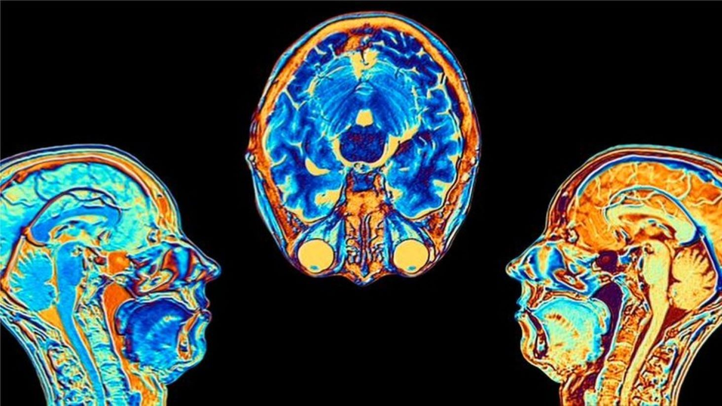
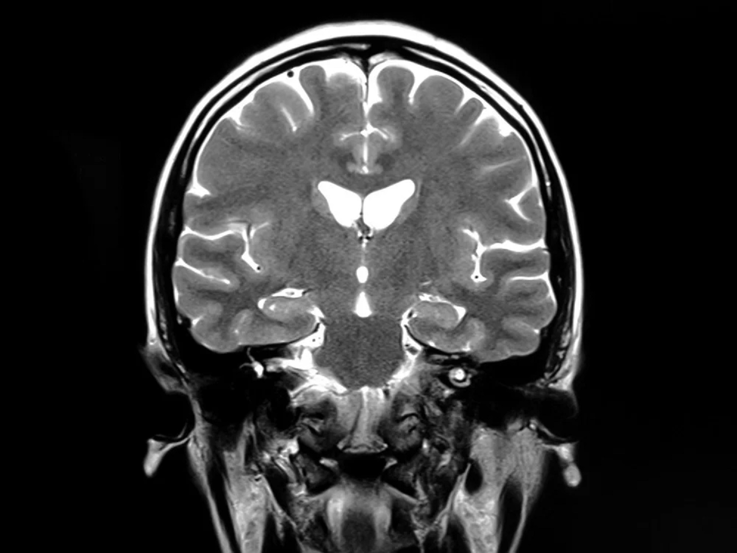
Previous studies examining lingering COVID-19 symptoms have primarily used MRI brain scans. While these studies have highlighted differences compared to healthy brains, these differences are not unique to COVID-19.
“As a doctor, it can be frustrating when patients report issues that MRI scans cannot explain.
This underscores the importance of exploring alternative imaging technologies to better understand the effects on the brain in patients with persistent COVID-19 symptoms,” explains Ida Blystad, a neuroradiologist at Linköping University Hospital and researcher associated with the Department of Health, Medicine and Caring Sciences at Linköping University, and the Centre for Medical Image Science and Visualization (CMIV).
In their recent study, researchers incorporated advanced diffusion MRI, a new imaging technique, to delve deeper into the brain’s white matter.
White matter primarily consists of nerve axons crucial for transmitting signals between different brain regions and the body.
“Diffusion MRI is highly sensitive, allowing us to detect changes in how nerve axons are organized.
This sensitivity prompted us to use diffusion MRI to investigate the impacts of COVID-19 on the brain, which may not be captured by other imaging methods,” says Deneb Boito, a doctoral student at the Department of Biomedical Engineering at Linköping University.
To grasp diffusion MRI, envision a bustling city at night where car lights illuminate major roads like strands of pearls.
Although the roads themselves aren’t visible, the movement of cars indicates their presence. Similarly, diffusion MRI enables doctors and researchers to understand the brain’s microscopic structure.
This technology exploits the movement of water molecules in the brain tissue, following paths of least resistance along neural pathways.
By measuring this movement, researchers can infer the brain’s neural pathway structure indirectly, akin to deducing the existence of a motorway from the movement of vehicles.
Diffusion MRI is widely used in healthcare for diagnosing stroke and planning brain surgeries.
In their study, researchers employed an advanced version of diffusion MRI to examine 16 male COVID-19 survivors from the Linköping COVID-19 Study (LinCos) at the Department of Rehabilitation Medicine in Linköping.
These individuals continued to experience symptoms seven months post-infection. They were compared with a group of healthy individuals without post-COVID symptoms who had not been hospitalized.
“The two groups exhibited differing brain white matter structures, potentially contributing to the neurological issues experienced by severe COVID-19 patients.
This aligns with other research indicating changes in white matter due to COVID-19. However, given our small sample size, we are cautious about drawing definitive conclusions.
This technology assesses brain microstructure rather than function. These findings underscore the need for advanced MRI techniques beyond conventional MRI to explore long-term COVID-19 effects on the brain,” notes Ida Blystad.
The researchers plan to delve deeper into several aspects. They observe variations in white matter across different brain regions but caution that it’s premature to interpret these variances.
A forthcoming study will explore whether diffusion MRI-detected changes correlate with brain activity and how different brain regions communicate via white matter in patients suffering from post-COVID fatigue.
Moreover, the researchers are keen to investigate temporal changes.
MRI captures a snapshot of the brain at a specific moment; thus, longitudinal studies are needed to determine if observed differences between groups persist over time or if they diminish.