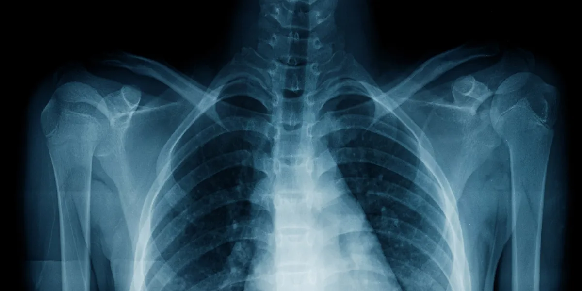
Chest X-rays are a common medical examination used globally to detect conditions such as tuberculosis and lung cancer. Despite their widespread use, these radiographs have not been able to assess lung functionality effectively. This limitation has been a significant gap in medical diagnostics, as lung function typically requires more invasive and patient-dependent tests.
Recent research published in The Lancet Digital Health has introduced a groundbreaking development in this area.
A team from Osaka Metropolitan University’s Graduate School of Medicine, led by Associate Professor Daiju Ueda and Professor Yukio Miki, has created an artificial intelligence model that can estimate lung function from chest X-rays with high precision. This innovation promises to transform how lung function is assessed, making it easier and more accurate.
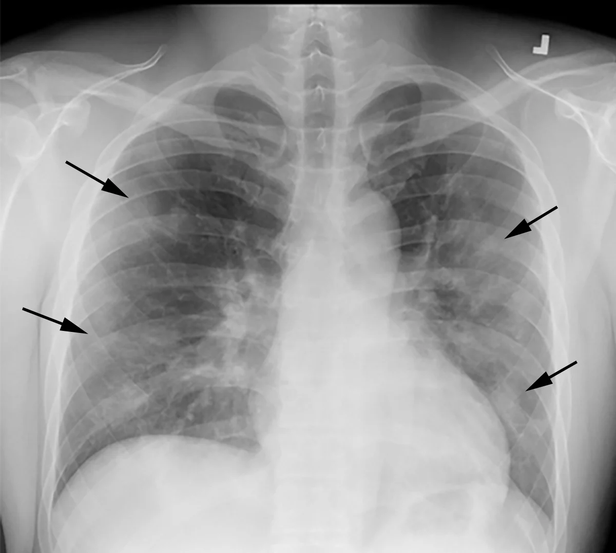
Traditionally, lung function is measured using spirometry, a process that requires patients to follow specific breathing instructions. This method can be challenging for certain populations, such as infants, people with dementia, or those who are physically incapacitated. The AI model developed by Professor Ueda’s team overcomes these limitations by using chest radiographs, which are far less demanding for patients.
The AI model was rigorously trained, validated, and tested using over 140,000 chest radiographs collected over nearly two decades. By comparing the AI’s estimates to actual spirometric data, the researchers fine-tuned the model to achieve a high level of accuracy.
The results were impressive, with the model demonstrating a Pearson’s correlation coefficient (r) of over 0.90, indicating strong agreement with traditional spirometry results.
This AI model holds significant promise for expanding pulmonary function assessment options, particularly for patients who struggle with conventional spirometry.
Professor Ueda emphasized the model’s potential to reduce patient burden and healthcare costs, highlighting the collaborative effort behind its development. If widely adopted, this AI-driven approach could revolutionize respiratory diagnostics by providing a non-invasive, reliable alternative to traditional lung function tests.